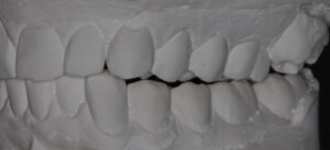
Making a splint for a patient with a scissors bite follows the same principles as in most other cases. A scissors bite is also called a buccal cross bite, wherein the maxillary molar is more than halfway across the mandibular molar; the lingual (tongue-side) cusp of the upper molar is touching the buccal (cheek-side) surface of the lower molar. A scissors bite looks like this:


The problem with a scissors bite is that the patient continually loses the sensation of contact with the left molars. As the eccentric functional pattern develops, more enamel is lost from this unusual bite relationship . . . and as more enamel is lost, the further the teeth erupt toward each other in this wayward direction.
A prominent feature in the patient with the R TMCC pattern is a loss of sensation of left molar contact. Placing a bite splint over the teeth that creates true second molar contact is difficult – particularly if the degree of buccal overlap is significant as in this case.
However, using the concept published in scientific papers by Trulsson et al, we can give the patient sensation of bite pressure with a tooth that doesn’t actually touch. Trulsson discovered that the input from nerve endings embedded in the tissue lining the tooth sockets are merged with as many as 2-4 adjacent teeth.
Here is how we solved this patient’s bite splint for scissors bite:

Splint creating normal bite sensation for upper cusp tip.

We were able to create overlapping tooth input so the splint for the scissors bite had the same sensation as if the teeth had a normal relationship, thus reducing the R TMCC patterning present.

